I am frequently asked: “Is my baby the right size?”, “When does my baby put on the most weight?” and “How big is my baby likely to be at birth?”
At each antenatal visit, I do ultrasound scanning of the unborn baby. There is no patient expense for this service. As well we give the patient at the first antenatal visit a free USB to store the images.
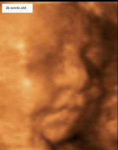
 Patients tell me how much they enjoy this service. They so often comment on how amazed they are by the imaging of their baby and particularly love to see the detail of their baby’s features and their baby moving. I get so much joy myself out of doing these scans. It is so good to see the joy of the parents and their bonding to their baby through seeing the ultrasound imaging. Patients are filled with such wonder and amazement. So often they say how much their baby’s size and appearance changes between visits. They can look back over past images stored on their USB
In early pregnancy, up to about 16 weeks, the estimate of the baby’s weight is based on the baby’s crown-rump (top of head to bottom) length measurement. The accuracy will be less at early gestations (less than 10 weeks) if the baby is moving excessively or is more curled up at the time of imaging.
 From about 16 weeks measurements of the baby’s head circumference, head width (biparietal diameter), abdominal circumference and femur (thigh bone) length are used to determine the estimated weight of the baby. These measurements are put into an algorithm developed by Dr Frank Hadlock and others, which is in the ultrasound scanner computer software. Use of Hadlock algorithm to determine foetal weight is a main steam approach used in many countries
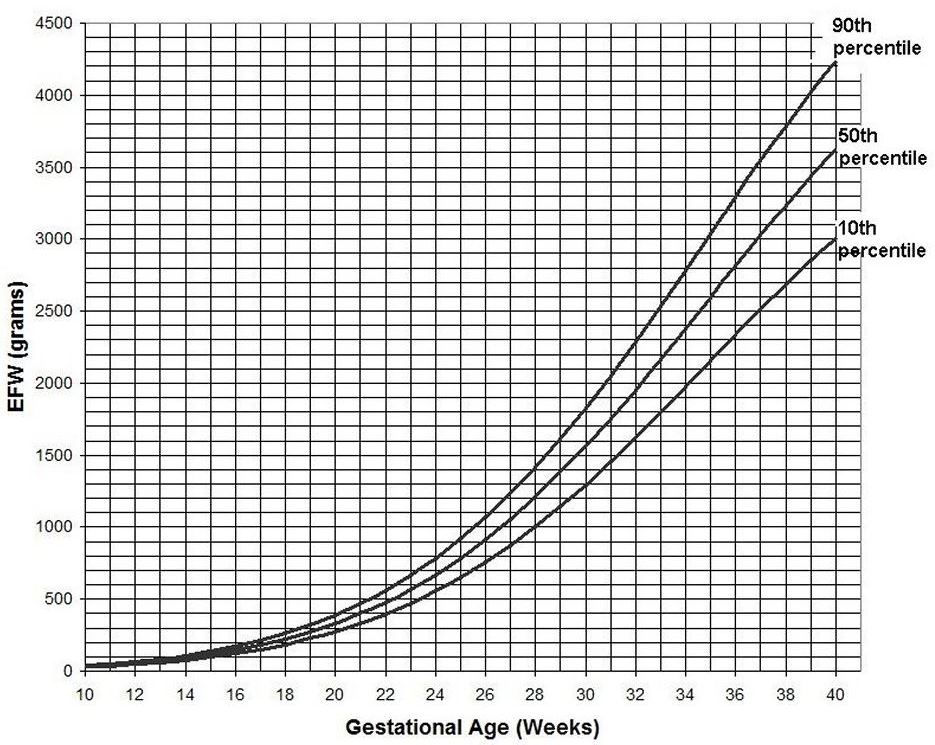
So you can understand how heavy a baby is at any number of weeks, baby’s growth at different gestations and what is the anticipated birth weight of baby I have included a chart derived from Dr Hadlock’s work. This has been sourced from the www.perinatology.com gestation calculator website and is reproduced with permission.

You can see from this chart that most of the weight gain happens at an increasing rate as the pregnancy advances.
The mean (average) weight at different weeks of pregnancy is shown in the chart above.
While the rate of increase of weight is the same for all races and ethnic backgrounds, the actual weight is based on a Caucasian person with a singleton pregnancy and without complications that would affect the baby’s size and growth.
I often get asked after 16 weeks: “What is the length of the baby now?” The answer is: “It is not possible to calculate this as the baby is too large and single imaging of the length of the baby on the screen is not possible”. A very approximate idea is to take the femur (thigh bone) length and multiply it by five. If you look at your own body your height is approximately five lengths of your thigh length.
Serial measurement of the baby at each antenatal visit is the most accurate way of assessing the baby’s growth. It is far more accurate than manually checking the height of the top (fundus) of the uterus or using a tape measure to measure the distance between the pubic bone and the fundus of the uterus. While ultrasound scanning is the most accurate way of determining the unborn baby’s size, it is not as accurate as weighing the baby, which is obviously not possible. The quoted error of ultrasound estimates is ± 15%. On a few occasions for various reasons, the error can be greater than this.
The baby’s growth being suboptimal or greater than usual raises the questions: “What is the reason for this?” and “What impact does this have on pregnancy care and delivery?” For instance, if the baby stops growing it implies the placenta is not functioning well and so early delivery is often needed to safeguard the baby’s wellbeing. There can be the opposite situation where the baby’s size is excessive. This may lead to a conversation about an earlier vaginal delivery to avoid the baby getting too big for the birth canal or even on a few occasions a Caesarean section rather than a vaginal delivery is recommended.
Serial ultrasound scanning of the baby gives patients so much joy and excitement. I personally get so much joy and it is a great pleasure to provide this service. Of more importance is that monitoring of foetal growth by serial ultrasound scans is an important part of antepartum care and provides the most accurate way of early detection of growth abnormalities and so helps to prevent foetal demise and to manage pregnancy complications more appropriately.
Hadlock FP, et al., In utero analysis of fetal growth: a sonographic weight standard. Radiology 1991 Oct;181(1):129-33.

